Anatomy Of Chest And Ribs / Chest Wall Lumps Rib Injury Clinic / Spiral ct of thoracic inlet.
byadmin•
0
Anatomy Of Chest And Ribs / Chest Wall Lumps Rib Injury Clinic / Spiral ct of thoracic inlet.. Finally, it describes the muscles that cause the motion in the chest wall. Pathology of the heart, mediastinum, lungs and pleura. These true ribs are also numerically known as the 1st, 2nd, 3rd, 4th, 5th, 6th, 7th, and the 8th ribs. The chest anatomy includes the pectoralis major, pectoralis minor and the serratus anterior. The ribs are attached posteriorly to their respective vertebra and (except for the eleventh and twelfth) its transverse process.
We hope you will use this picture in the study and helping chest and abdominal cavities with some organs removed. The anatomical structure of the 24 ribs in the human body is complex because of the irregular shape and different lengths of each rib. As part of the bony thorax, the ribs protect the internal thoracic organs. These true ribs are also numerically known as the 1st, 2nd, 3rd, 4th, 5th, 6th, 7th, and the 8th ribs. And as you might guess from the word major, it makes up the majority of the chest muscle mass.
Thoracic Chest And Back Skeletal Skeleton Anatomy Featuring The Ribs Stock Photo Alamy from c8.alamy.com To carry out the unique functions performed by. This is a commonly performed procedure and is necessary in ibrahim, af and darwish: It can help you understand our world more detailed and specific. As with all parts of the body, the anatomy and physiology of the chest wall are intimately intertwined. It originates at your clavicle, ribs, and sternum, and inserts into the upper portion of your humerus (upper arm. The embryologic and anatomic basis of modern surgery. The rib cage also anchors the bones of the head, neck, shoulders, and arms to the trunk of the body. Spiral ct of thoracic inlet.
We hope you will use this picture in the study and helping chest and abdominal cavities with some organs removed.
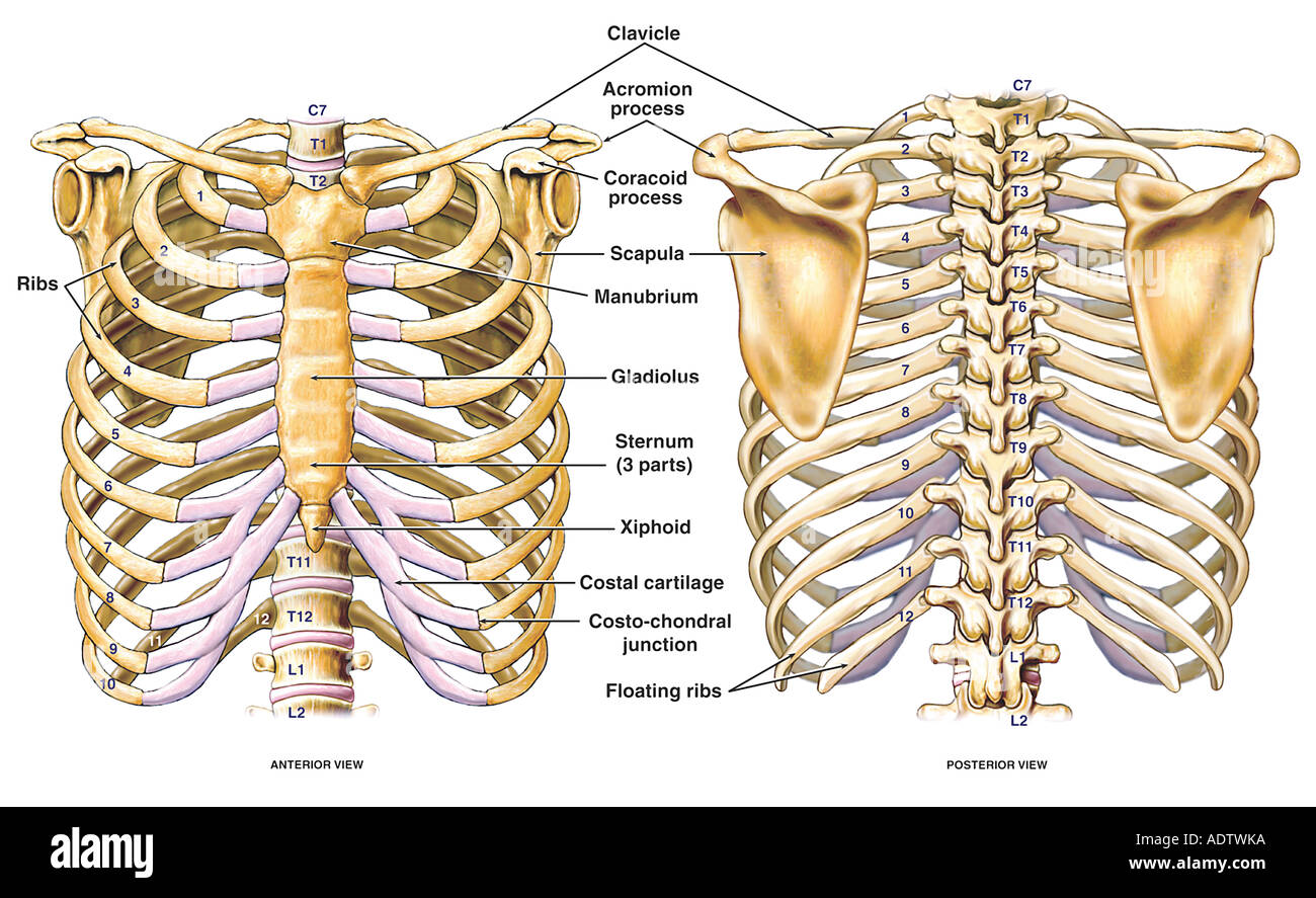
The bones of the chest and upper back combine to form the strong protective rib cage around the vital thoracic organs such as the heart and. The embryologic and anatomic basis of modern surgery. These true ribs are also numerically known as the 1st, 2nd, 3rd, 4th, 5th, 6th, 7th, and the 8th ribs. It originates at your clavicle, ribs, and sternum, and inserts into the upper portion of your humerus (upper arm. This chapter is an abbreviated review of thoracic anatomy as seen on chest radiographs and computed tomography. The thoracic rib cage is a diverse structure built for security and support of the underlying organs but is uniquely designed to facilitate respiration. This type of ct scan uses a lower radiation level than a conventional. On the standard left lateral chest radiograph, the right ribs are projected behind the left and appear. Related posts of chest bone anatomy. The purpose of this study was to explore the effect of. The anatomical structure of the 24 ribs in the human body is complex because of the irregular shape and different lengths of each rib. As with all parts of the body, the anatomy and physiology of the chest wall are intimately intertwined. As part of the bony thorax, the ribs protect the internal thoracic organs.
The length of each space corresponds to that of the adjacent ribs and their cartilages; This chapter is an abbreviated review of thoracic anatomy as seen on chest radiographs and computed tomography. They also have a role in ventilation; On the standard left lateral chest radiograph, the right ribs are projected behind the left and appear. This type of ct scan uses a lower radiation level than a conventional.
1 Thoracic Wall Radiology Key from i1.wp.com It originates at your clavicle, ribs, and sternum, and inserts into the upper portion of your humerus (upper arm. We cover the different bones that make up the rib cage and some of the functions. Chest bone, ribs, lung, heart, xiphoid process. The rib cage surrounds the lungs and the heart, serving as an important means of bony protection for these vital organs. Surface anatomy of anterior chest wall. It discusses the specific anatomy of the ribs and costal cartilages, along with the sternum. Anatomy is to physiology as geography is to history: The spectrum of these rare anomalies includes unilateral absence, absence of cartilage, separation of cartilage and rib, combined skandalakis' surgical anatomy:
Respiratory muscle training strengthen the function of the respiratory muscles to improve your patient's overall.
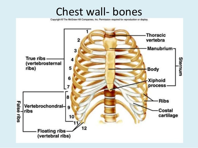
How these parts interrelate through joints is described also. Insert contains images of a typical rib and the first rib. How these parts interrelate through joints is described also. Ribs eight to ten are the false ribs and are connected to the sternum indirectly via the cartilage of the rib clinical notes. It discusses the specific anatomy of the ribs and costal cartilages, along with the sternum. The rib cage also anchors the bones of the head, neck, shoulders, and arms to the trunk of the body. Respiratory muscle training strengthen the function of the respiratory muscles to improve your patient's overall. The rib cage surrounds the lungs and the heart, serving as an important means of bony protection for these vital organs. Chest blunt trauma (cbt) and the resultant rib fractures often lead to thoracic collapse. This is a commonly performed procedure and is necessary in ibrahim, af and darwish: The bones of the chest and upper back combine to form the strong protective rib cage around the vital thoracic organs such as the heart and. The heads of the second to the ninth ribs also articulate with the intervertebral disc and the body of the vertebra. Finally, it describes the muscles that cause the motion in the chest wall.
Posteriorly, the heads of the ribs interdigitate with the vertebrae and are numbered according to the inferior vertebra. The bones of the chest and upper back combine to form the strong protective rib cage around the vital thoracic organs such as the heart and. And as you might guess from the word major, it makes up the majority of the chest muscle mass. Pathology of the heart, mediastinum, lungs and pleura. O bones—spine, ribs, clavicles, scapulae, humeri.
Chest Bone Anatomy Anatomy Drawing Diagram from image.slidesharecdn.com The costotransverse ligaments in human: The spectrum of these rare anomalies includes unilateral absence, absence of cartilage, separation of cartilage and rib, combined skandalakis' surgical anatomy: And as you might guess from the word major, it makes up the majority of the chest muscle mass. The bones of the chest and upper back combine to form the strong protective rib cage around the vital thoracic organs such as the heart and. Related online courses on physioplus. Each rib wraps around the lung and descends approximately 3 to 5 inches. Ribs are divided into two basic groups the true ribs consist of 8 ribs, each on the left and right sides of the chest wall. What are the features of ribs?
This is a commonly performed procedure and is necessary in ibrahim, af and darwish:
This chapter is an abbreviated review of thoracic anatomy as seen on chest radiographs and computed tomography. Insert contains images of a typical rib and the first rib. Finally, it describes the muscles that cause the motion in the chest wall. Ribs eight to ten are the false ribs and are connected to the sternum indirectly via the cartilage of the rib clinical notes. Spiral ct of thoracic inlet. Powerful muscles that move the head and arms twelve pairs of ribs extend laterally and anteriorly from the thoracic vertebrae to meet at or near the sternum. We hope you will use this picture in the study and helping chest and abdominal cavities with some organs removed. Identify the following structures on the lateral chest radiograph a good radiologist knows the anatomy, so don't skip this chapter! As with all parts of the body, the anatomy and physiology of the chest wall are intimately intertwined. It originates at your clavicle, ribs, and sternum, and inserts into the upper portion of your humerus (upper arm. It can help you understand our world more detailed and specific. O bones—spine, ribs, clavicles, scapulae, humeri. It discusses the specific anatomy of the ribs and costal cartilages, along with the sternum.
How these parts interrelate through joints is described also anatomy of chest. ■ identify the basic anatomy seen on a chest radiograph.